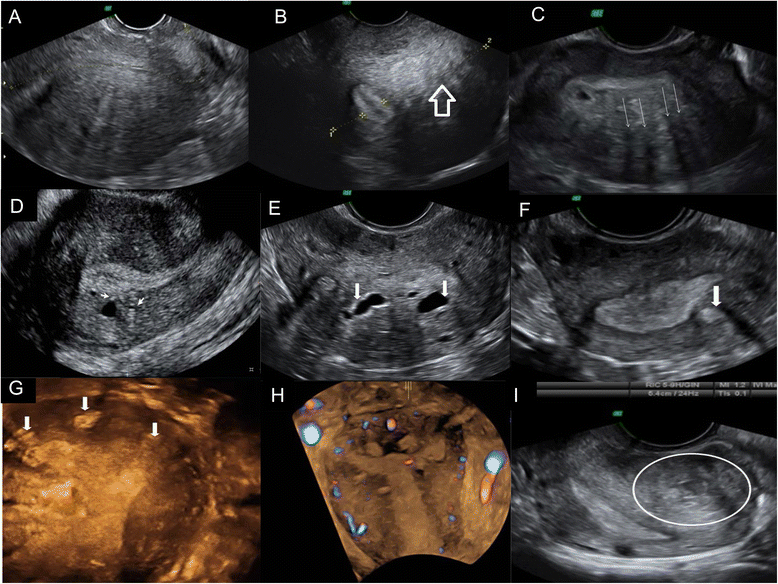
Fig. 1

Ultrasonographic diagnostic criteria for adnomyosis. a Globulous aspect of the uterus. b Uterine asymmetry. Longitudinal section of a retroverted uterus, where the posterior uterine wall is clearly thicker than the anterior wall. c Heterogeneous myometrial texture. Transversal section of the uterus at the fundus level, where hypoechoic areas with radial pattern can be seen (arrows). d Linear striations. In this sagital section of an anteverted uterus thin hyperecogenic lines cross the myometrial thickness, visible from the endometrial-myometrial interphase. e Intramyometrial cysts. Transversal section of the uterus at the fundus level with sonoluscent images distributed in posterior wall of the myometrium. f and g, h Hyperechogenic nodules. Transversal (f) and coronal (g, h) sections of the uterus at the fundus level where hyperechogenic Intramyometrial areas can be observed (arrows). i Adenomyoma. Longitudinal section of a retroverted uterus with heterogeneous nodular mass lacking well-defined margins in the posterior wall